|
|
SAINT ...PHONIE | |||
BREATH AND CRANIO-SACRAL RHYTHMS |
||||
|
In the literature, the sacrocranial rhythms refer to the palpation of the waves of the patient by his therapist. But it is also possible for each one of us to connect to the listening of our own rhythms and to interact with them.
Breathing in the therapeutist - patient interactionWhen the therapist connects himself to the “primary respiration” rhythm (CRI) of his patient, it appears frequently that the breathing of the therapist fits automatically in phase with the CRI of its patient. It is also often noticed that the osteopathic treatment is facilitated when the patient is aware of his own breathing during the treatment.
Breathing and listening to our own rhythmsWhen we are listening at our own CRI (4-9 cycles/minute), and if we let our breathing rock us on his rhythm, we reach another part of ourselves. Connecting to this “ancestral” part of ourselves creates a major wellbeing and a feeling of reliance with the nature which surrounds us. If we analyze the variations of frequencies of the cardiac rhythm associated in this state, we observe (fig. 7) :
Fig 7: variations of cardiac rhythm HRV (in light blue), of breathing (dark blue), HF (white) and LF (brown). When we analyze the size of the peak of the cardiac pulsations, we can see that the amplitude of these peaks follows breathing in a synchronous way: the size of the peak increases during expiration and decreases during inspiration (fig. 8). We observe a phase shift between the end of the inspiration and the maximum of cardiac pulsations (it is the opposite of figure 5).
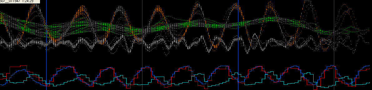
Fig 8: variations of cardiac rhythm HRV (in light blue), of breathing (dark blue), HF (white), LF (brown), VLF (in green) When we continue our internalization process, listen to our own MID-TIDE (1-3 cycles/minute) and let our breathing rock us on his rhythm, it is the waveband corresponding to the VLF which is activated (fig. 9). In this case, the minimum of VLF wave corresponds to one of the minima of LF wave and to one of maximum of the HF wave. The three wavebands are in “harmony”.
When we analyze the amplitude (size of the peaks) of the cardiac pulsations, wee notice that the size of these peaks always follows breathing in a synchronous way: the size of the peak increases during expiration and decreases during inspiration (fig. 10). Compared to figures 5 and 8, the shift between the end of the inspiration and the maximum of cardiac pulsations increases: and it is during expiration that the cardiac rhythm increases.
Fig 10: variations of cardiac rhythm HRV (in light blue), of breathing (dark blue), HF (white), LF (brown), VLF (in green)
|
|||
| Tous
droits réservés - © P. BOTTE 2010 - compteur statistique
:
|