|
|
SAINT ...PHONIE | |||
CARDIO-BREATHING SYSTEM ANALYSIS AND OSTEOPATHY |
||||
|
VARIOUS RHYTHMS PALPED BY THE OSTEOPATHSThe different rhythmsThe osteopaths working with the cranio-sacral therapy learn how to palpate and interact with some types of rhythms generated by the fluctuations of the cerebro-spinal fluid. They distinguish three types of rhythms:
Most used and more known, the CRI or “primary respiration” is used “to treat” (to return a better mobility, to give again a harmony of movement) the bones, the muscles and the organs which have a certain fixity. There is a disconcerting agreement between the rhythms palpated by the osteopaths and the frequencies extracted from the analysis of cardiac variability:
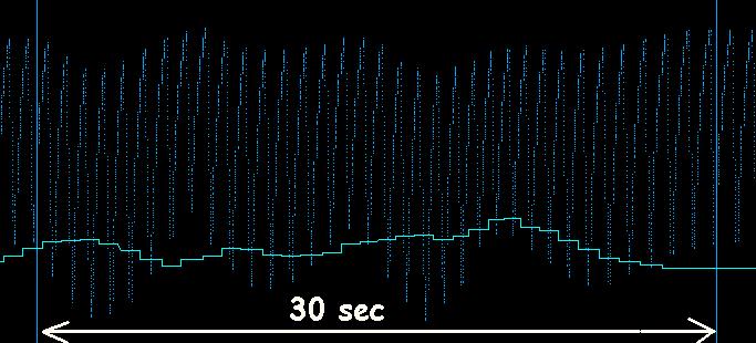
Experimental validation of the palpated rhythmsAt the end of the 19th century, Traube, Hering and Meyer highlighted a fluctuation of the pressure of the cardiac impulses which one calls Traube-Hering-Meyer oscillation (fig. 4). This fluctuation may still be present in absence of breathing.
Fig 4.: variations of the cardiac pulsations recorded by an auricle laser Doppler system highlighting the Traube-Hering-Meyer oscillations (dark blue curve). The light blue curve represents the monitoring of the variations of the cardiac rhythm (HRV). It is observed that the variation of the Traube-Hering-Meyer wave does not follow the variations of heart rate (HRV). When one connects the recording of Traube-Hering-Meyer wave and Osteopathic palpation, we obtain a good correlation between flexion and extensions identified by manual palpation and fluctuations of the Traube-Hering-Meyer wave.
Recording of the cardiac Traube-Hering-Mayer wave (light blue curve). In yellow, the recording of the manual palpation. In dark blue, monitoring of respiration. One can observe a good correlation between the upper part of the light blue curve and the manual palpation of the sacro-cranial wave (yellow curve). The lower part of the cardiac signal (light blue) follows respiration (dark blue).
Several authors connected the oscillations of Traube Hering-Meyer with the palpation of the “primary respiration” rhythm (CRI):
Concerning the slower rhythm (mid-tide), it was shown that the intracranial pressure followed oscillations (called B waves) having a periodicity ranging between 30 seconds and 2 minutes (ANDERS EKLUND and Al (reference O5) and Mikael Edsbagge et al. (reference O6)).
|
|||
| Tous
droits réservés - © P. BOTTE 2010 - compteur statistique
:
|